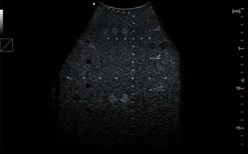
TFM (Total Focusing Method) is an ultrasonic array technique used to achieve full focusing at every point within a region of interest.It is also a data processing method that uses FMC (Full Matrix Capture) to acquire data for synthetic focusing,which involves image reconstruction within the region of interest (pixel frame) to focus on a large number of points forming a grid.
1.Ultra-wideband Uniform Beam Emission
Full-focus imaging breaks through the beam shape limitations of traditional fixed-point focusing, enabling intelligent dynamic beam control over the entire region.It delivers consistent high resolution across the tissue area without sacrificing frame rate.
2.Stronger and More Uniform Energy
Traditional ultrasound energy is concentrated near the selected focal point and attenuates rapidly away from the focus.Full-focus imaging overcomes this limitation via real-time correlated energy-modulated transmission, achieving uniform energy distribution throughout the imaging field.
3.Superior Penetration
The resolution of traditional ultrasound images is limited by the focal position and propagation speed of acoustic waves in tissue.Based on an RF metadata platform, full-focus imaging applies holographic correction and beam optimization to full-field RF metadata with large-scale real-time variations,delivering exceptional detail resolution and excellent penetration at high frequency and high resolution, thereby producing high-quality images.
Full-focus imaging transcends the technical limitations of conventional ultrasound imaging platforms and solves the problem of out-of-focus blur in traditional ultrasound.It provides clinicians with superior image quality, more accurate diagnostic tools, and a smarter clinical application experience.
Ultrafast Imaging
Ultrafast ultrasound imaging breaks through the frame rate limitations of conventional ultrasound, enabling the capture of higher-resolution, sharper images at a frame rate 100 times faster than traditional imaging.This will drive further advancements in prevention, diagnosis, and treatment monitoring.
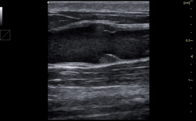
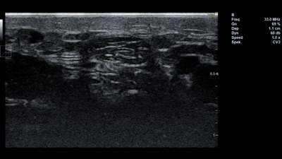
CMUT Technology
Utilizing CMUT (Capacitive Micromachined Ultrasonic Transducer) semiconductor ultrasound technology, this system elevates image spatial resolution to micrometer level high definition imaging, providing clinicians with clear 2D images for observing and diagnosing the epidermis, dermis, subcutaneous tissue, and skin appendages.Rich and sensitive color Doppler flow imaging is also available to assess subcutaneous blood flow and the status of skin tumors.